Small bowel obstruction is a common surgical emergency. Adhesions from previous surgery cause most cases in the developed world; hernias, tumours, Crohn's strictures and gallstone ileus account for most of the rest.
Clinical presentation
Colicky central abdominal pain, vomiting, distension and absolute constipation. Constant pain, fever, tachycardia or peritonism suggest strangulation.
Radiographic features
Abdominal radiograph
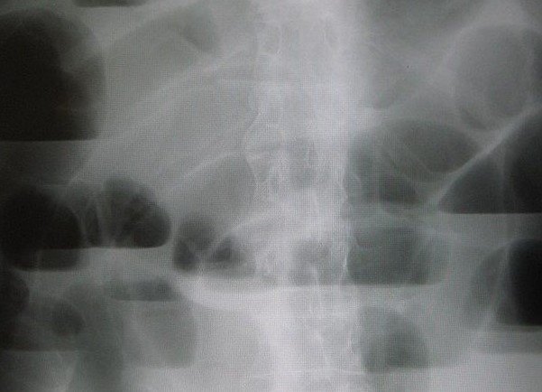
- Dilated small bowel loops (more than about 3 cm) lying centrally
- Valvulae conniventes crossing the full width of the bowel
- Multiple air–fluid levels on an erect or decubitus film
- Little or no gas in the colon
CT
CT confirms obstruction, finds the transition point between dilated and collapsed bowel, identifies the cause and looks for ischaemia: reduced wall enhancement, mesenteric oedema, free fluid or pneumatosis. The small bowel faeces sign often lies just proximal to the transition point.
Treatment and prognosis
Adhesional obstruction without signs of ischaemia is usually managed with nasogastric decompression and fluids; water-soluble contrast can be both diagnostic and therapeutic. Strangulation, closed-loop obstruction or failure to settle require surgery.
Differential diagnosis
- Paralytic ileus (dilatation of both small and large bowel, no transition point)
- Large bowel obstruction
- Gastroenteritis
Cases and figures
References
- Ten Broek RPG, Krielen P, Di Saverio S, et al. Bologna guidelines for diagnosis and management of adhesive small bowel obstruction (ASBO): 2017 update of the evidence-based guidelines from the World Society of Emergency Surgery ASBO working group. World J Emerg Surg. 2018;13:24.
Cite this entry
Small bowel obstruction. Radflow Encyclopedia. Last revised 9 November 2026. https://radflow.cc/encyclopedia/small-bowel-obstruction (accessed 15 November 2026).
Related entries
Educational reference for healthcare professionals and students. It is not medical advice and does not replace clinical judgement or local guidelines.