Lobar pneumonia describes infective consolidation that spreads through the alveoli of a lobe, typically bounded by a fissure. Streptococcus pneumoniae is the classic cause, though many organisms can produce the pattern.
Clinical presentation
Fever, productive cough, pleuritic pain and breathlessness, often with focal crackles or bronchial breathing over the affected lobe. Older patients may present only with confusion or falls.
Pathology
Exudate fills the air spaces while the bronchi remain aerated. Because infection spreads through the pores of Kohn, the consolidation is homogeneous and stops at a fissure.
Radiographic features
Plain radiograph
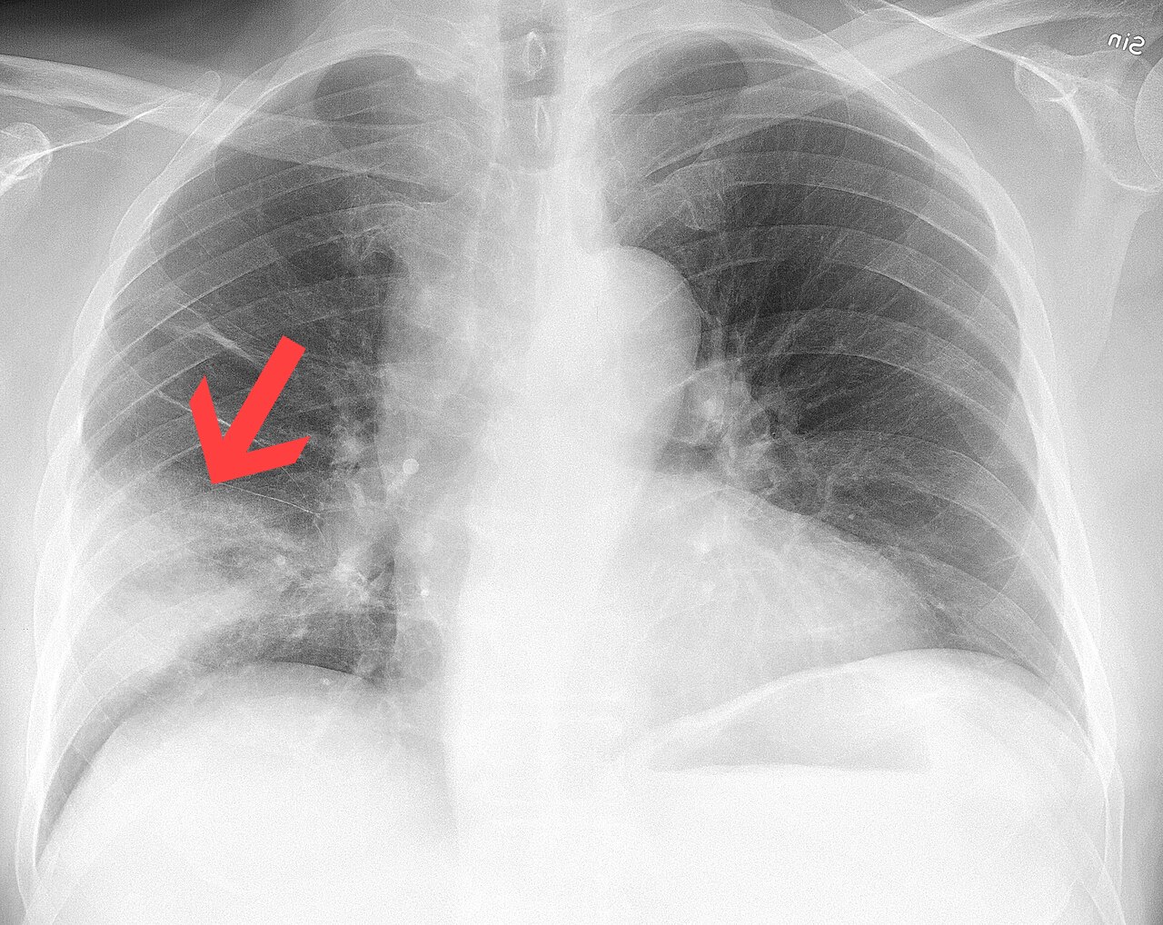
- Homogeneous opacity confined to a lobe, often with a sharp fissural margin
- Air bronchograms: dark, branching airways within the white consolidation
- The silhouette sign localises the lobe — for example, loss of the right heart border with right middle lobe disease
- Volume is usually preserved, unlike collapse
CT
CT is not needed for straightforward cases but helps when there is suspected cavitation, empyema or an obstructing lesion.
Treatment and prognosis
Antibiotics are guided by severity scores and local guidance. Radiographic clearing lags behind clinical improvement and may take six weeks or more; a follow-up film is commonly advised in smokers and older patients to exclude an underlying tumour.
Differential diagnosis
- Pulmonary haemorrhage or oedema
- Lobar collapse (look for volume loss)
- Organising pneumonia
- Mucinous adenocarcinoma, especially if non-resolving
Cases and figures
References
- Metlay JP, Waterer GW, Long AC, et al. Diagnosis and treatment of adults with community-acquired pneumonia. An official clinical practice guideline of the American Thoracic Society and Infectious Diseases Society of America. Am J Respir Crit Care Med. 2019;200(7):e45–e67.
Cite this entry
Lobar pneumonia. Radflow Encyclopedia. Last revised 13 November 2026. https://radflow.cc/encyclopedia/lobar-pneumonia (accessed 15 November 2026).
Related entries
Educational reference for healthcare professionals and students. It is not medical advice and does not replace clinical judgement or local guidelines.