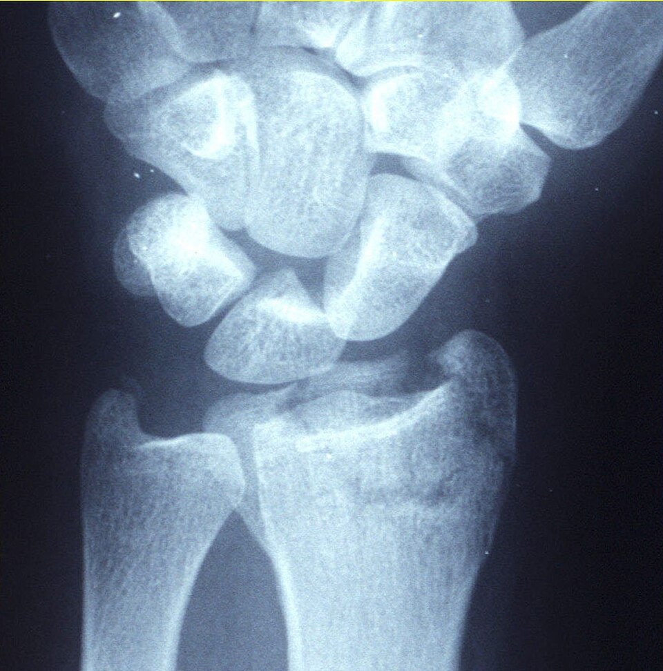
Distal radius fractures are among the most common fractures seen in emergency departments. They occur in a bimodal pattern: high-energy injuries in the young and low-energy falls in older people with osteoporosis.
Classification
- Colles: dorsal angulation and displacement of the distal fragment
- Smith: volar angulation (a reverse Colles)
- Barton: intra-articular fracture–dislocation of the dorsal or volar rim
- Chauffeur: radial styloid fracture
Radiographic features
Plain radiograph
PA and lateral views are assessed for:
- Radial inclination (normally about 22°) and radial height (about 11 mm)
- Volar tilt on the lateral view (normally about 11°)
- Intra-articular extension and step-off
- Associated ulnar styloid fracture and distal radioulnar joint injury
CT
Helps plan surgery for comminuted intra-articular fractures.
Treatment and prognosis
Undisplaced or acceptably reduced fractures are treated in a cast. Unstable or intra-articular fractures may need K-wires or volar plate fixation. Older patients often do well functionally even with imperfect alignment.
Practical points
Always assess the scaphoid and the carpal alignment on wrist films; associated injuries are easy to miss.
Cases and figures
References
- Medoff RJ. Essential radiographic evaluation for distal radius fractures. Hand Clin. 2005;21(3):279–288.
Cite this entry
Distal radius fracture. Radflow Encyclopedia. Last revised 1 November 2026. https://radflow.cc/encyclopedia/distal-radius-fracture (accessed 15 November 2026).
Related entries
Educational reference for healthcare professionals and students. It is not medical advice and does not replace clinical judgement or local guidelines.