Case 1013
Staghorn calculusWhat is the diagnosis?
 X-ray
1 / 1
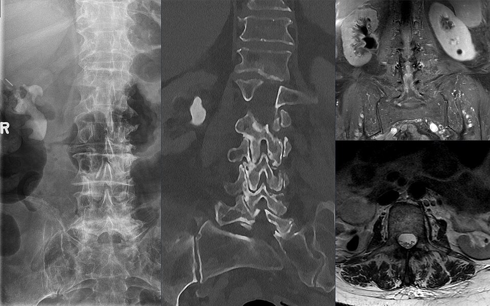
Composite: plain radiograph, coronal CT and MRI of the same patient.
Image: Hellerhoff, CC BY-SA 4.0 — sourceAnswer hidden. Review the images and history, decide on your diagnosis, then reveal.
Findings
- A large, branching radiopaque calculus projects over the right kidney on the plain film
- Coronal CT confirms a dense stone filling the right renal pelvis and extending into the calyces
- On MRI the stone is a signal void within the collecting system
Case discussion
A staghorn calculus fills the renal pelvis and branches into the calyces. Most are struvite stones linked to infection with urease-producing organisms, so recurrent urinary infection is a typical presentation.
Untreated, they can destroy the kidney. Percutaneous nephrolithotomy is the usual treatment; infected, obstructed systems need urgent drainage.