Case 1009
Extradural hematomaWhat is the diagnosis?
 CT
1 / 1
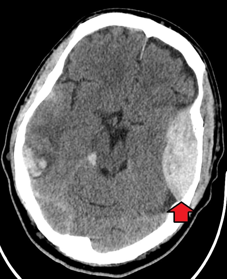
Axial non-contrast CT of the head (arrow from the original source).
Image: James Heilman, MD, CC BY-SA 4.0 — sourceAnswer hidden. Review the images and history, decide on your diagnosis, then reveal.
Findings
- Biconvex (lentiform) hyperdense extra-axial collection over the left hemisphere (arrow)
- Mass effect with effacement of the adjacent sulci
- Additional hyperdense foci in the brain parenchyma, consistent with contusions
Case discussion
A lens-shaped collection that does not cross suture lines is an extradural hematoma, usually from a tear of the middle meningeal artery associated with a temporal bone fracture. Check bone windows for the fracture.
Deterioration can be rapid. Large hematomas or neurological decline need urgent neurosurgical evacuation; outcomes are often good when this happens quickly.