Case 1007
Acute appendicitisWhat is the diagnosis?
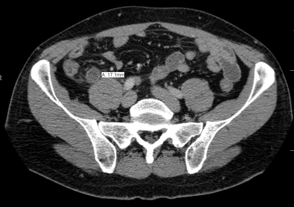 CT
1 / 2
Axial contrast-enhanced CT of the pelvis; the calliper measures the appendix (17.1 mm).
Image: James Heilman, MD, CC BY-SA 3.0 — sourceAnswer hidden. Review the images and history, decide on your diagnosis, then reveal.
Findings
- Dilated appendix measuring about 17 mm (normal under 6 mm)
- Thickened, enhancing wall with surrounding fat stranding
- Ultrasound shows a thick-walled, tubular, non-compressible appendix
Case discussion
An appendix over 6 mm with wall thickening and periappendiceal inflammation is diagnostic of acute appendicitis in the right clinical setting.
Ultrasound is first-line in children and pregnancy; CT is preferred in most adults when the diagnosis is uncertain. Look for complications — perforation, abscess and extraluminal gas — that change management.
Encyclopedia
More cases like this.
All cases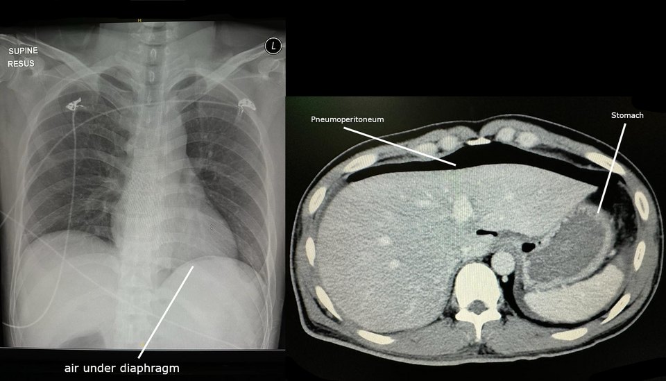 2
Pneumoperitoneum
Sudden severe epigastric pain, now generalised. Rigid abdomen on examination.
 2
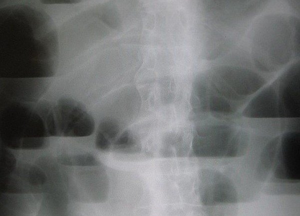
Small bowel obstruction
Colicky central abdominal pain, vomiting and distension. Previous laparotomy.
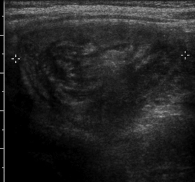
Ileocolic intussusception
Episodes of inconsolable crying with drawing up of the legs, and vomiting.